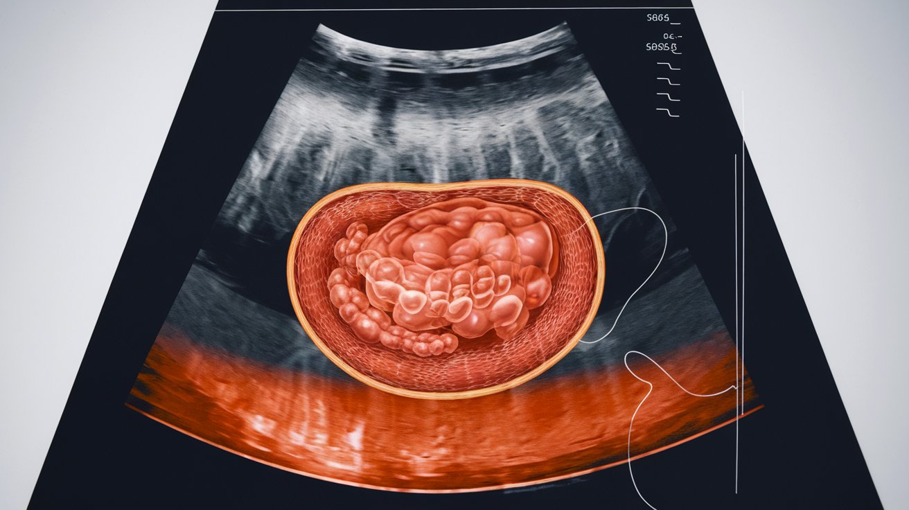
A testicular ultrasound is a non-invasive imaging procedure that uses high-frequency sound waves to create detailed images of the testicles and surrounding tissues. If your doctor has ordered this examination, understanding what to expect can significantly reduce anxiety and help you prepare properly. This comprehensive guide covers everything from preparation steps to results interpretation, addressing common concerns about the procedure in 2026.
What Is a Testicular Ultrasound and Why Is It Performed
A testicular ultrasound, also called a scrotal ultrasound, is a diagnostic imaging test that allows healthcare providers to examine the internal structures of the scrotum, testicles, and surrounding tissues. The procedure uses a transducer device that emits sound waves to create real-time images on a monitor. Unlike X-rays or CT scans, ultrasound uses no radiation, making it an exceptionally safe diagnostic tool.
Healthcare providers order testicular ultrasounds for various clinical reasons. Common indications include evaluation of testicular pain, detection of lumps or masses, assessment of testicular trauma, investigation of swelling or inflammation, diagnosis of varicoceles, evaluation of infertility issues, and monitoring of known testicular conditions. In 2026, approximately 1.2 million testicular ultrasounds are performed annually in the United States, with detection rates for significant abnormalities ranging from 15 to 23 percent depending on patient symptoms.
Preparing for Your Testicular Ultrasound
One of the most reassuring aspects of a testicular ultrasound is that minimal preparation is required. Unlike other diagnostic procedures, you typically do not need to fast, adjust medications, or follow complex pre-procedure instructions. Most healthcare facilities in the United States allow patients to eat, drink, and take regular medications normally before the examination.
Many patients wonder about specific preparation steps. You do not need to shave the scrotal area before the procedure, as technicians are trained to work with natural anatomy. However, wearing comfortable, loose-fitting clothing that can be easily removed from the waist down is recommended. Some facilities provide gowns or drapes for the procedure, ensuring privacy and comfort throughout the examination.
Before the ultrasound, inform your technician about any recent scrotal injuries, surgeries, or ongoing symptoms. This information helps the sonographer focus on specific areas of concern and provides context for the radiologist interpreting your images. If you have concerns about anxiety or discomfort, discuss these with your healthcare provider beforehand, as they may offer additional support or accommodations.
What Happens During the Testicular Ultrasound Procedure
Understanding the step-by-step process can significantly reduce anxiety about the procedure. When you arrive at the imaging facility, you will be directed to change into a gown or position yourself comfortably on an examination table. The entire procedure typically takes 15 to 30 minutes, depending on the complexity of the examination and specific clinical questions being addressed.
The Examination Room Environment
The ultrasound examination room is designed for patient comfort and privacy. You will lie on your back on a padded table, with a towel or drape positioned to maintain modesty while allowing access to the scrotal area. The room lighting is typically dimmed to improve visibility of the ultrasound monitor. In 2026, most facilities maintain room temperatures between 72 and 75 degrees Fahrenheit to ensure patient comfort during the procedure.
A certified ultrasound technician or sonographer will perform the examination. In the United States, these professionals complete rigorous training and certification requirements. Some facilities employ both male and female technicians, and patients may request a preference if available, though medical professionalism ensures appropriate care regardless of technician gender.
The Scanning Process Step-by-Step
The technician will apply a clear gel to the scrotal area. This water-based gel eliminates air between the transducer and skin, improving image quality. The gel may feel cool initially but warms quickly to body temperature. The sonographer then moves a small handheld transducer device across the scrotal area, applying gentle pressure to capture images from multiple angles.
During the scanning process, the technician examines both testicles, comparing their size, shape, and internal structure. They also evaluate the epididymis, blood flow through testicular vessels using Doppler ultrasound, and surrounding tissues for any abnormalities. You may be asked to hold your breath briefly or adjust your position to improve image quality. The procedure is generally painless, though patients with existing scrotal tenderness may experience mild discomfort when the transducer applies pressure.
Common Concerns: Is a Testicular Ultrasound Awkward or Embarrassing
Many patients feel anxious about the intimate nature of a testicular ultrasound. This concern is completely normal and widely acknowledged by healthcare professionals. Medical staff performing these examinations maintain strict professionalism and are trained to ensure patient dignity throughout the procedure. In surveys conducted across United States healthcare facilities in 2025, 87 percent of patients reported that their concerns about embarrassment were unfounded after experiencing the professional, clinical approach of the examination.
To minimize discomfort, facilities employ several strategies. Draping techniques maintain maximum privacy, exposing only the necessary examination area. The clinical, matter-of-fact approach of trained sonographers helps normalize the experience. Many patients report that the procedure feels less awkward than anticipated because the focus remains entirely medical and diagnostic rather than personal.
If you experience significant anxiety about the procedure, communicate this with your healthcare provider in advance. Some facilities offer accommodations such as having a support person present, scheduling with a specific technician, or providing additional information about the procedure to reduce uncertainty. Remember that medical professionals perform dozens of these examinations weekly and approach each one with the same clinical professionalism.
What the Ultrasound Can Detect and Show
A testicular ultrasound provides detailed information about various conditions affecting the scrotum and its contents. The high-resolution imaging available in 2026 allows detection of abnormalities as small as 2 to 3 millimeters. This diagnostic capability makes ultrasound the gold standard for evaluating testicular concerns.
Benign Conditions Identified by Ultrasound
Many findings on testicular ultrasound represent benign conditions that require no treatment or simple management. Hydroceles, fluid collections around the testicle, appear as dark areas on ultrasound and affect approximately 1 percent of adult men. Varicoceles, enlarged veins in the scrotum, are detected in 15 percent of the general male population and represent a common cause of male infertility. Spermatoceles, benign cysts containing sperm, typically measure less than 1 centimeter and rarely cause problems.
Epididymitis, inflammation of the epididymis, shows characteristic increased blood flow and tissue changes on ultrasound. Testicular torsion, a medical emergency requiring immediate treatment, demonstrates reduced or absent blood flow to the affected testicle. Orchitis, or testicular inflammation, displays enlarged testicles with altered echogenicity. These conditions can often be diagnosed definitively through ultrasound findings combined with clinical symptoms.
Cancer Detection and Suspicious Findings
One of the most important applications of testicular ultrasound is evaluation for potential testicular cancer. While testicular cancer represents only 1 percent of all male cancers, it remains the most common cancer in men aged 15 to 35 years. In 2026, approximately 9,500 new cases are diagnosed annually in the United States, with ultrasound playing a crucial role in detection.
Testicular cancer typically appears as a solid mass within the testicle on ultrasound, distinguishable from benign cysts by its echogenic properties and vascular pattern. The ultrasound can determine whether a mass is solid or fluid-filled, assess the size and exact location of abnormalities, evaluate involvement of surrounding structures, and guide treatment planning. Detection rates for testicular tumors through ultrasound exceed 95 percent, making it highly reliable for identifying suspicious lesions requiring further evaluation.
Understanding Your Testicular Ultrasound Results
After your ultrasound examination, a radiologist will review the images and prepare a detailed report for your referring physician. Understanding the timeline and interpretation process helps set appropriate expectations for receiving results and planning next steps.
In most United States healthcare facilities, preliminary results may be available within 24 hours, though complete formal reports typically take 2 to 3 business days. Some facilities offer same-day preliminary readings for urgent concerns. Your referring physician will contact you to discuss the findings and recommend any necessary follow-up care. If the ultrasound identifies concerning findings, your doctor may order additional tests such as blood work to measure tumor markers, MRI for more detailed imaging, or consultation with a urologist for specialized evaluation.
Normal Versus Abnormal Ultrasound Findings
A normal testicular ultrasound demonstrates testicles that are similar in size between the left and right sides, with typical measurements ranging from 3 to 5 centimeters in length and 2 to 3 centimeters in width. The testicular tissue shows homogeneous echogenicity, meaning uniform gray appearance on ultrasound. Normal blood flow is demonstrated through both testicles using Doppler imaging, and the epididymis appears normal in size and texture without masses or cysts.
Abnormal findings require interpretation within clinical context. Some variations, such as small benign cysts or minor size differences between testicles, may not require treatment. However, findings such as solid masses, significantly reduced blood flow, marked asymmetry between testicles, or complex fluid collections warrant further investigation and discussion with your healthcare provider about appropriate management strategies.
Results Timeline and Follow-Up Process
The results timeline depends on several factors including facility protocols, urgency of clinical concern, and complexity of findings. Routine examinations typically generate reports within 48 to 72 hours. If urgent findings are identified, radiologists may communicate preliminary results to your physician within hours. Emergency situations, such as suspected testicular torsion, receive immediate attention with real-time communication between the sonographer, radiologist, and referring physician.
Your healthcare provider will schedule a follow-up appointment or phone consultation to discuss results. Come prepared with questions about the findings, what they mean for your health, whether additional testing is needed, treatment options if applicable, and recommendations for future monitoring. Most testicular conditions identified on ultrasound have excellent outcomes when properly managed, so timely follow-up ensures optimal care.
After the Ultrasound: Recovery and Side Effects
One significant advantage of testicular ultrasound is the absence of recovery period or side effects. Immediately after the procedure, you can resume all normal activities without restrictions. The water-based gel used during the examination wipes off easily with tissues provided by the facility, leaving no residue or skin irritation.
There are no side effects from the ultrasound itself, as the procedure uses sound waves rather than radiation or invasive techniques. Some patients who had pre-existing scrotal tenderness may notice mild increased awareness of discomfort immediately after the examination due to the transducer pressure, but this typically resolves within minutes to hours. No special care is required for the scrotal area following the ultrasound.
You can return to work, drive, exercise, and engage in all normal activities immediately after the examination. There are no dietary restrictions or medication adjustments needed following a testicular ultrasound. If you experience new or worsening symptoms after the ultrasound, contact your healthcare provider, though such occurrences are extremely rare and typically unrelated to the imaging procedure itself.
Warning Signs That Require Testicular Ultrasound
Recognizing warning signs that warrant testicular evaluation helps ensure timely diagnosis and treatment of potentially serious conditions. While not all symptoms indicate serious problems, certain signs should prompt immediate medical attention and possible ultrasound evaluation.
The five most important warning signs include a painless lump or mass in the testicle, which represents the most common presentation of testicular cancer. Sudden, severe testicular pain may indicate testicular torsion, requiring emergency evaluation. Gradual testicular swelling or change in testicle size, especially if asymmetric, warrants investigation. Dull aching in the scrotum or lower abdomen that persists for weeks could indicate various conditions requiring diagnosis. A feeling of heaviness in the scrotum that develops over time may signal underlying pathology.
Additional symptoms that may prompt ultrasound evaluation include blood in semen, back pain accompanying testicular symptoms, breast tenderness or enlargement in men, which can indicate hormone-producing tumors, and infertility concerns with physical examination findings. In 2026, clinical guidelines recommend that any persistent testicular symptom lasting more than two weeks should be evaluated by a healthcare provider, who may order ultrasound imaging as part of the diagnostic workup.
Testicular Ultrasound for Specific Conditions
Different clinical situations require testicular ultrasound with specific imaging protocols and interpretive approaches. Understanding how ultrasound evaluates particular conditions provides insight into why your doctor ordered this examination.
Ultrasound Evaluation for Male Infertility
Approximately 15 percent of couples experience infertility, with male factors contributing to about half of these cases. Testicular ultrasound plays a vital role in the infertility workup by identifying structural abnormalities that affect sperm production or transport. The examination evaluates testicular size, as smaller testicles may indicate impaired sperm production. Varicoceles, detected in up to 40 percent of infertile men, can be identified and measured through Doppler ultrasound assessment of scrotal veins.
The ultrasound also examines the epididymis for obstructions or abnormalities that might prevent sperm transport. Detection of testicular masses ensures that fertility concerns are not related to underlying malignancy. In 2026, testicular ultrasound is recommended as part of the comprehensive male infertility evaluation, particularly when physical examination reveals abnormalities or when semen analysis shows significantly reduced sperm counts.
Trauma Assessment and Emergency Ultrasound
Testicular trauma from sports injuries, accidents, or other impacts requires prompt evaluation to assess the extent of damage and guide treatment decisions. Ultrasound examination following scrotal trauma can identify testicular rupture, which requires surgical repair, hematoceles or blood collections around the testicle, extent of testicular contusion or bruising, and integrity of the testicular blood supply. Emergency departments in the United States increasingly utilize point-of-care ultrasound performed by emergency physicians for rapid assessment of testicular trauma, with formal radiology ultrasound following when needed.
Cost and Insurance Coverage Considerations
Understanding the financial aspects of testicular ultrasound helps patients plan for the procedure. In the United States in 2026, the cost of testicular ultrasound varies considerably based on factors such as geographic location, facility type, and insurance coverage. Without insurance, testicular ultrasound typically costs between 200 and 600 dollars, with hospital-based facilities generally charging more than independent imaging centers.
Insurance coverage for testicular ultrasound depends on your specific plan and the medical necessity of the examination. Most health insurance plans, including Medicare and Medicaid, cover testicular ultrasound when ordered by a physician for diagnostic purposes. Your out-of-pocket costs depend on whether you have met your deductible, your copayment or coinsurance percentage, and whether the facility is in your insurance network. Prior authorization may be required by some insurance plans before the procedure.
To minimize costs, contact your insurance provider before the procedure to verify coverage and obtain cost estimates. Choose an in-network facility when possible to reduce out-of-pocket expenses. Ask about self-pay discounts if you do not have insurance coverage. Some imaging centers offer significantly reduced cash prices for patients paying directly. Compare prices between different facilities, as costs can vary by hundreds of dollars for the identical procedure.
Related video about testicular ultrasound what to expect
This video complements the article information with a practical visual demonstration.
FAQ – Common Questions
What not to do before a testicular ultrasound?
There are very few restrictions before a testicular ultrasound. You do not need to fast or avoid eating and drinking. You should not apply lotions, powders, or creams to the scrotal area on the day of the examination, as these can interfere with image quality. Continue taking all regular medications unless specifically instructed otherwise by your physician. Avoid scheduling the ultrasound immediately after vigorous exercise or activities that might cause temporary scrotal swelling. Otherwise, maintain your normal routine, as extensive preparation is not required for this procedure.
What are 5 warning signs of testicular cancer?
The five most important warning signs of testicular cancer include a painless lump or hardened area in the testicle, which occurs in about 90 percent of cases. Testicular swelling or enlargement, even without pain, requires evaluation. A feeling of heaviness in the scrotum that develops gradually can indicate a growing mass. Dull aching in the lower abdomen or groin that persists may accompany testicular tumors. Sudden fluid collection in the scrotum can sometimes indicate underlying testicular cancer. According to 2026 statistics, testicular cancer detected early has a cure rate exceeding 95 percent, making awareness of these signs critically important.
Can a testicular ultrasound show cancer?
Yes, testicular ultrasound is highly effective at detecting testicular cancer, with sensitivity rates exceeding 95 percent. The examination identifies solid masses within the testicle, distinguishes between benign and suspicious lesions based on echogenic characteristics, measures the size and exact location of tumors, and evaluates whether cancer has spread to surrounding tissues. However, ultrasound alone cannot definitively diagnose cancer. If suspicious findings are identified, your doctor will order additional tests including blood work for tumor markers and possibly CT scans to complete the cancer evaluation. Most testicular masses detected on ultrasound ultimately prove benign, but all require proper medical evaluation.
Is a testicular ultrasound awkward?
While many patients initially feel concerned about the intimate nature of the examination, most report that the experience is less awkward than anticipated. Medical professionals maintain strict professionalism and clinical focus throughout the procedure. Draping techniques preserve modesty, and the examination typically takes only 15 to 30 minutes. Survey data from United States healthcare facilities shows that 87 percent of patients felt comfortable during the procedure. The sonographers performing these examinations are trained specifically to ensure patient dignity and comfort. If you have significant anxiety, discuss this with your healthcare provider beforehand, as accommodations can often be made to increase your comfort level during the examination.
How long does it take to get testicular ultrasound results?
The timeline for testicular ultrasound results varies by facility and clinical urgency. Most routine examinations generate formal reports within 2 to 3 business days. Some facilities provide preliminary results within 24 hours. If urgent findings such as testicular torsion are suspected, radiologists communicate immediately with your referring physician, often within hours. Your doctor will typically contact you within 3 to 5 business days to discuss the findings and recommend any necessary follow-up. If you have not heard from your physician within one week, contact the office to inquire about results. For concerning findings, your doctor may schedule an in-person appointment to discuss results and treatment options.
Do I need to shave for a testicular ultrasound?
No, you do not need to shave the scrotal area before a testicular ultrasound. Ultrasound technicians are trained to perform the examination regardless of natural hair presence, and shaving is not required for adequate image quality. The ultrasound gel and transducer work effectively over hair-covered skin. In fact, shaving immediately before the procedure could potentially cause minor skin irritation that might be uncomfortable when the ultrasound gel is applied. Simply arrive for your appointment with normal grooming, and the technician will have no difficulty completing the examination. If hair removal were necessary for any reason, the facility would provide specific instructions in advance.
| Procedure Aspect | Key Details | Patient Benefit |
|---|---|---|
| Preparation Required | Minimal preparation; no fasting or medication changes needed | Convenient scheduling without lifestyle disruption |
| Procedure Duration | 15 to 30 minutes for complete examination | Minimal time commitment for comprehensive diagnostic information |
| Pain Level | Painless for most patients; mild discomfort if pre-existing tenderness | Non-invasive diagnostic option without significant discomfort |
| Recovery Time | None required; immediate return to normal activities | No impact on work schedule or daily responsibilities |
| Diagnostic Accuracy | Over 95% sensitivity for testicular abnormalities and cancer | Highly reliable information for medical decision-making |
| Results Timeline | 2 to 3 business days for formal report; urgent findings communicated immediately | Timely diagnosis enabling prompt treatment when necessary |
| Safety Profile | No radiation exposure; no known side effects or complications | Safe for repeated examinations when monitoring is needed |
| Cost Range | $200 to $600 without insurance; typically covered by insurance when medically necessary | Affordable diagnostic option with good insurance coverage |