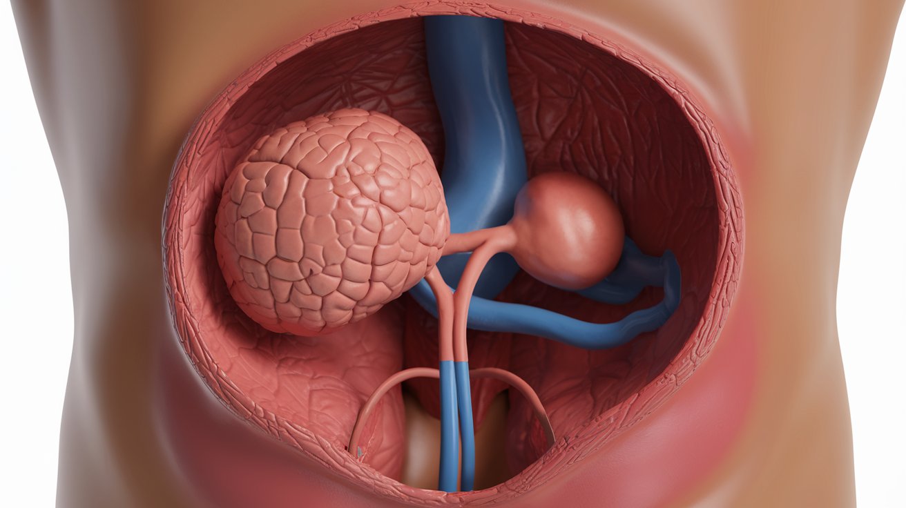
The prostate gland is located in the male pelvis, positioned directly below the bladder and in front of the rectum. This walnut-sized gland surrounds the first part of the urethra, the tube that carries urine from the bladder through the penis. Understanding the precise anatomical location of the prostate is essential for recognizing symptoms of prostate problems and understanding diagnostic procedures used by urologists across the United States.
Precise Anatomical Position of the Prostate Gland
The prostate gland occupies a specific position within the male pelvis, making it accessible for clinical examination while protecting it within the pelvic cavity. This small but crucial organ sits in the space between several important anatomic structures. The gland measures approximately 3 centimeters in length and 4 centimeters in width in healthy adult males, though these dimensions vary with age and health status. Its strategic location allows it to perform its reproductive functions while remaining protected by surrounding pelvic bones and tissues.
The prostate is positioned centrally in the pelvis, not on the left or right side. It rests on the pelvic floor muscles called the levator ani, which provide support from below. Above the prostate sits the bladder, with the connection point between these two organs forming the bladder neck. The prostatic urethra runs through the center of the gland, creating a passage for both urine and seminal fluid. This central positioning is why prostate enlargement can significantly impact urinary function, as the growing tissue compresses the urethra from all sides.
Relationship to Surrounding Anatomical Structures
Understanding the prostate’s relationship to nearby organs helps explain both its function and the symptoms that arise when problems develop. The rectum lies immediately behind the prostate, separated only by a thin layer of tissue called Denonvilliers’ fascia. This proximity is why digital rectal examination remains the primary physical method for assessing prostate size, texture, and abnormalities. Urologists can feel the posterior surface of the gland through the anterior rectal wall during routine examinations.
The seminal vesicles attach to the prostate’s posterior-superior surface, just behind where the bladder meets the gland. These paired structures contribute fluid to semen and empty their contents through ducts that pass through the prostate tissue. The vas deferens, the tubes carrying sperm from the testicles, also pass into the prostate to join with the seminal vesicle ducts. Below and around the prostate run crucial neurovascular bundles containing nerves responsible for erectile function, which is why prostate surgery can sometimes affect sexual function. The membranous urethra exits the prostate at its apex (lowest point) and passes through the pelvic floor muscles before continuing as the bulbar urethra.
Internal Structure and Zones of the Prostate
The prostate is not a uniform organ but consists of distinct zones with different cellular compositions and disease patterns. The most clinically significant division recognizes four main zones: the peripheral zone, central zone, transitional zone, and anterior fibromuscular stroma. The peripheral zone comprises approximately 70% of glandular tissue in young men and is located primarily at the back and sides of the gland. This zone is where approximately 70-80% of prostate cancers originate, making it the focus of biopsy procedures and screening examinations.
The transitional zone surrounds the urethra and accounts for only 5-10% of the prostate in young adults. However, this zone is where benign prostatic hyperplasia (BPH) develops, causing the gland to enlarge with age. As the transitional zone grows, it can compress the urethra and cause urinary symptoms affecting millions of American men over age 50. The central zone makes up about 25% of the gland and rarely develops cancer. The anterior fibromuscular stroma contains no glandular tissue but consists of muscle and fibrous tissue that provides structural support. Understanding these anatomical zones helps physicians target biopsies and plan surgical approaches for different prostate conditions.
Primary Functions of the Prostate Gland
The prostate performs several essential functions related to male reproduction and urinary control. Its primary role involves producing prostatic fluid, which constitutes approximately 30% of seminal fluid volume. This milky, slightly alkaline fluid contains enzymes, citric acid, zinc, and prostate-specific antigen (PSA). The prostatic secretions help nourish and protect sperm after ejaculation, neutralizing the acidic environment of the vaginal tract and providing nutrients that sustain sperm viability during their journey to fertilize an egg.
During ejaculation, smooth muscle fibers within the prostate contract to propel semen through the urethra and out of the penis. The gland also contains a sphincter mechanism at the bladder neck that prevents retrograde ejaculation (semen flowing backward into the bladder) and helps maintain urinary continence. The prostate produces PSA enzyme that liquefies semen after ejaculation, changing it from a gel-like consistency to a liquid state that allows sperm to swim freely. In 2026, researchers continue exploring additional biochemical functions of prostatic secretions, including their role in immune function and potential antimicrobial properties that protect the urinary tract.
How to Locate Your Prostate: Physical Examination
Many men wonder if they can feel their own prostate and where to locate it. The prostate cannot be felt from the outside through the abdominal wall or perineum due to its internal pelvic position. However, the posterior surface of the prostate can be palpated through the anterior wall of the rectum during a digital rectal exam (DRE). Healthcare providers insert a lubricated, gloved finger approximately 3-4 inches into the rectum to reach the prostate’s posterior surface.
A normal prostate feels smooth, firm, and roughly walnut-sized during examination. The gland should be symmetrical with a slight median groove dividing the right and left lobes. Abnormal findings include hard nodules, asymmetry, tenderness, or significant enlargement. While self-examination is technically possible, medical professionals recommend professional evaluation for accurate assessment. The angle and technique required make self-examination difficult and unreliable. American Urological Association guidelines recommend that men discuss prostate screening with their healthcare provider starting at age 45-50, depending on risk factors including family history and ethnicity.
Blood Supply and Nervous System Connections
The prostate receives its blood supply primarily from the inferior vesical artery, a branch of the internal iliac artery. Additional arterial contributions come from the middle rectal artery and internal pudendal artery. These vessels branch extensively within the prostatic tissue, creating a rich vascular network necessary for the gland’s metabolic functions. The venous drainage flows through the prostatic venous plexus, also called the plexus of Santorini, which surrounds the gland anteriorly and laterally. This venous system connects to the internal iliac veins and provides a potential route for cancer spread in advanced prostate malignancies.
The innervation of the prostate involves both autonomic and somatic nervous systems. Sympathetic nerve fibers from the hypogastric plexus and parasympathetic fibers from the pelvic splanchnic nerves regulate prostatic secretion and smooth muscle contraction. The cavernous nerves, which are crucial for erectile function, run along both sides of the prostate in neurovascular bundles. These nerve bundles typically travel between 2-5 millimeters lateral to the prostatic capsule. Damage to these nerves during prostatectomy procedures can result in erectile dysfunction, which is why nerve-sparing surgical techniques have become standard practice. In 2026, advanced imaging and robotic surgery allow surgeons to better preserve these delicate nerve structures during prostate cancer treatment.
Common Prostate Problems and Location Symptoms
The prostate’s location makes it vulnerable to several age-related conditions that affect urinary and sexual function. Three primary disorders affect the gland: benign prostatic hyperplasia (BPH), prostatitis, and prostate cancer. Each condition produces symptoms related to the gland’s anatomical position and its relationship to the urethra and bladder. Understanding how location influences symptoms helps men recognize when to seek medical evaluation.
Benign Prostatic Hyperplasia and Urinary Symptoms
Benign prostatic hyperplasia represents non-cancerous enlargement of the transitional zone that affects over 50% of men in their 60s and up to 90% of men over 80 in the United States. As the transitional zone grows inward, it compresses the prostatic urethra, creating obstruction to urine flow. Early symptoms include difficulty starting urination, weak stream, frequent urination especially at night (nocturia), and sensation of incomplete bladder emptying. The location-based compression explains why these symptoms worsen gradually as the gland continues enlarging. Treatment options in 2026 range from medications that relax prostatic smooth muscle or shrink gland tissue to minimally invasive procedures like transurethral resection (TURP) or newer techniques using steam, laser, or water vapor ablation.
Prostatitis and Pelvic Pain Syndromes
Prostatitis involves inflammation or infection of the prostate gland that can occur at any age but most commonly affects men under 50. The condition causes pain in the perineum (area between scrotum and anus), lower back, penis, or testicles due to the prostate’s location and nerve connections. Men may experience painful urination, difficulty urinating, or pain during ejaculation. Acute bacterial prostatitis causes severe symptoms with fever and requires immediate antibiotic treatment. Chronic prostatitis/chronic pelvic pain syndrome is more common but harder to treat, often requiring extended therapy with medications, physical therapy, and lifestyle modifications. The deep pelvic location makes this condition challenging to diagnose and treat effectively.
Prostate Cancer Location and Detection
Prostate cancer predominantly develops in the peripheral zone at the back and sides of the gland, which explains why digital rectal examination can detect suspicious nodules. This location also makes the peripheral zone accessible to transrectal ultrasound-guided biopsy procedures. In 2026, approximately 288,000 new cases of prostate cancer are diagnosed annually in the United States, making it the most common non-skin cancer in American men. Early-stage prostate cancer typically causes no symptoms due to its peripheral location away from the urethra. Advanced disease may cause urinary obstruction, blood in urine or semen, bone pain from metastases, or erectile dysfunction when cancer invades the neurovascular bundles.
First Warning Signs of Prostate Problems
Recognizing early warning signs of prostate disease enables prompt medical evaluation and better treatment outcomes. The five most significant warning signs include changes in urinary frequency, difficulty initiating or maintaining urine flow, urgent need to urinate, blood in urine or semen, and pain in the pelvic region. Increased nighttime urination (waking 2 or more times) often represents the earliest symptom men notice, as the enlarged gland gradually restricts urine flow from the bladder.
Additional warning signs include weak or interrupted urine stream, straining to urinate, sensation of incomplete bladder emptying, and dribbling at the end of urination. Pain or burning during urination may indicate infection or inflammation. Erectile dysfunction or painful ejaculation can signal prostate problems affecting the surrounding neurovascular structures. Blood in the urine (hematuria) or semen (hemospermia) requires immediate evaluation, though these symptoms more commonly result from benign conditions than cancer. Lower back pain, hip pain, or bone pain in men over 50 with known prostate issues may indicate advanced cancer with bone metastases. The American Cancer Society recommends that men with any persistent urinary symptoms or concerning signs consult a urologist for comprehensive evaluation including PSA testing and physical examination.
Diagnostic Procedures and Prostate Imaging
Modern diagnostic techniques exploit the prostate’s anatomical position to visualize and assess the gland through multiple approaches. The digital rectal examination remains the foundational physical assessment method, allowing urologists to evaluate size, symmetry, consistency, and the presence of nodules. Blood tests measuring PSA levels provide biochemical assessment, with elevated levels potentially indicating BPH, prostatitis, or cancer. In 2026, age-adjusted PSA reference ranges and PSA density calculations improve the specificity of this screening tool.
Transrectal ultrasound (TRUS) uses sound waves transmitted through a probe inserted into the rectum to create detailed images of prostate anatomy. This technique guides needle biopsies to sample suspicious areas within the gland. Multiparametric MRI has revolutionized prostate imaging by providing superior soft tissue contrast that identifies suspicious lesions before biopsy. MRI fusion biopsy techniques combine MRI images with real-time ultrasound for targeted sampling of abnormal areas in the peripheral zone. Computed tomography (CT) and bone scans assess cancer spread beyond the prostate. Urodynamic testing evaluates how prostate enlargement affects bladder function and urine flow. These complementary diagnostic approaches leverage the gland’s accessible location while providing comprehensive assessment of structure, function, and disease.
Prostate Location in Females: Anatomical Equivalent
Women do not have a prostate gland in the traditional sense, but they possess anatomically equivalent structures called Skene’s glands or paraurethral glands. These small structures located on the anterior wall of the vagina near the urethra are considered homologous to the male prostate because they develop from the same embryonic tissue. The Skene’s glands produce small amounts of fluid released during sexual arousal and may contribute to female ejaculation in some women.
Research has demonstrated that Skene’s glands contain tissue similar to prostatic tissue, including cells that produce PSA in small quantities. These glands can develop conditions analogous to male prostate problems, including cysts, infections, and rarely, adenocarcinoma. The term “female prostate” is sometimes used in medical literature, though these structures are much smaller than the male prostate and do not surround the urethra in the same manner. Understanding this anatomical relationship helps explain why PSA is not exclusively a male biomarker, though levels in women remain significantly lower. In 2026, ongoing research continues exploring the functional roles of female paraurethral glands and their relationship to urinary and sexual health.
Sexual Function After Prostate Surgery
One of the most common concerns men have involves whether erections are possible after prostate removal or treatment. The answer depends on several factors including the type of procedure, surgical technique, patient age, and pre-existing erectile function. The neurovascular bundles running alongside the prostate contain nerves essential for erectile function. When these nerves are preserved during surgery, many men retain the ability to achieve erections, though recovery may take 6-24 months.
Radical prostatectomy performed using nerve-sparing techniques preserves erectile function in 50-70% of appropriate candidates, with better outcomes in younger men and those with strong pre-operative function. Robotic-assisted laparoscopic prostatectomy has improved nerve preservation rates compared to open surgery by allowing better visualization and precision. However, even with perfect nerve preservation, some degree of erectile dysfunction is common immediately after surgery due to neuropraxia (temporary nerve injury). Rehabilitation protocols using PDE5 inhibitors (sildenafil, tadalafil), vacuum devices, or penile injections help preserve erectile tissue during recovery. Radiation therapy affects erectile function more gradually, with dysfunction developing in 30-50% of men over several years. In 2026, advanced surgical techniques, better patient selection, and comprehensive rehabilitation programs have significantly improved sexual outcomes for men requiring prostate cancer treatment.
Related video about where is prostate located
This video complements the article information with a practical visual demonstration.
Key Questions and Answers
Where exactly is the prostate located in the male body?
The prostate is located in the male pelvis, directly below the bladder and in front of the rectum. It surrounds the first portion of the urethra as it exits the bladder. The gland sits centrally in the pelvis, not on the left or right side, and rests on the pelvic floor muscles. This strategic position allows it to add secretions to semen while making it accessible for examination through the rectum. The prostate measures approximately 3 centimeters long and 4 centimeters wide in healthy adults.
What are the first signs of a prostate problem?
The first signs of prostate problems typically involve changes in urination patterns. Early warning signs include increased frequency of urination especially at night, difficulty starting urination, weak urine stream, urgent need to urinate, and sensation of incomplete bladder emptying. Some men notice straining to urinate or dribbling after urination. Pain or burning during urination, blood in urine or semen, pelvic discomfort, or erectile dysfunction may also indicate prostate issues. These symptoms result from the prostate’s location surrounding the urethra, where enlargement or inflammation compresses the urinary passage. Any persistent urinary symptoms warrant medical evaluation.
Can you feel your prostate from the outside?
No, you cannot feel the prostate from the outside of your body through the abdomen or perineum due to its deep pelvic location. The only way to physically feel the prostate is through the rectum during a digital rectal examination. A healthcare provider can palpate the posterior surface of the prostate by inserting a gloved, lubricated finger approximately 3-4 inches into the rectum. The gland sits against the anterior rectal wall, making it accessible for examination. A normal prostate feels smooth, firm, and symmetrical, roughly the size of a walnut, with a slight groove in the middle.
What are the 5 warning signs of an enlarged prostate?
The five primary warning signs of an enlarged prostate (BPH) include: 1) Frequent urination, especially waking multiple times at night; 2) Difficulty starting urination or weak urine stream; 3) Urgent, sudden need to urinate; 4) Sensation of incomplete bladder emptying; and 5) Dribbling at the end of urination or straining to urinate. These symptoms occur because the enlarging transitional zone of the prostate compresses the urethra running through its center. Over 50% of men in their 60s and 90% over age 80 experience some degree of prostate enlargement. Treatment options range from medications to minimally invasive procedures depending on symptom severity.
Can you get an erection without a prostate?
Yes, erections are possible without a prostate, though erectile function may be affected by the surgery required to remove it. The prostate itself does not directly cause erections, but the neurovascular bundles containing erection nerves run alongside the prostate. When these nerves are preserved during prostatectomy, 50-70% of appropriate candidates retain erectile function, though recovery takes 6-24 months. Younger men with good pre-operative function have better outcomes. Even when nerves are damaged, erectile function may be restored using medications, vacuum devices, or penile implants. The prostate’s role is producing fluid for semen, not controlling erections.
Is the prostate in the anus or rectum?
No, the prostate is not located in the anus or rectum. The prostate sits in the pelvic cavity in front of the rectum, separated by a thin layer of tissue. However, because the rectum passes directly behind the prostate, the gland’s posterior surface can be felt through the anterior rectal wall during a digital rectal examination. This anatomical relationship makes rectal examination the primary physical method for assessing prostate size and detecting abnormalities. The prostate is positioned between the bladder above and the pelvic floor below, with the rectum behind it and the pubic bone in front.
| Anatomical Aspect | Key Details | Clinical Significance |
|---|---|---|
| Location | Below bladder, in front of rectum, surrounds urethra | Accessible for examination; enlargement affects urination |
| Size | 3cm long, 4cm wide (walnut-sized in healthy adults) | Increases with age; monitoring helps detect abnormal growth |
| Primary Function | Produces 30% of seminal fluid with PSA enzyme | Essential for reproduction; PSA used as screening marker |
| Zones | Peripheral (70%), transitional (10%), central (25%) | Different zones prone to specific diseases (cancer vs BPH) |
| Blood Supply | Inferior vesical artery with rich venous plexus | Important for surgical planning; potential cancer spread route |
| Nerve Bundles | Cavernous nerves run 2-5mm lateral to gland | Preservation crucial for maintaining erectile function post-surgery |